
PRF, A-PRF, i-PRF Webinar — Professor Choukroun
Good evening, dear colleagues!
First of all, I would like to thank PlusSkyBuy for giving me the opportunity to conduct this webinar.
As you know, it is currently midnight in Nice, France, but it is a great pleasure for me to share my scientific and clinical experience with you. In this webinar, we will discuss PRF — platelet-rich fibrin.

Growth factors are used in everyday surgical dental practice. However, if we want to improve our clinical outcomes, we must use the right materials and the right techniques.
To achieve our goals, we need cells, hormones, proteins, and growth factors. These help us achieve good healing. However, the main goal we are trying to achieve is new angiogenesis.
We usually use platelet concentrate techniques, in which centrifugation is used to increase the concentration of platelets. This was the original concept behind the development of regenerative products.
Today, we have three types of protocols:
- PRP — platelet-rich plasma; anticoagulants are used.
- PRGF — plasma rich in growth factors; anticoagulants are used.
- PRF — platelet-rich fibrin; no anticoagulants are used.
What are the differences between them? Should we use anticoagulants or not? Why should we avoid anticoagulants? By the way, be sure to read about how to choose the correct tubes here.
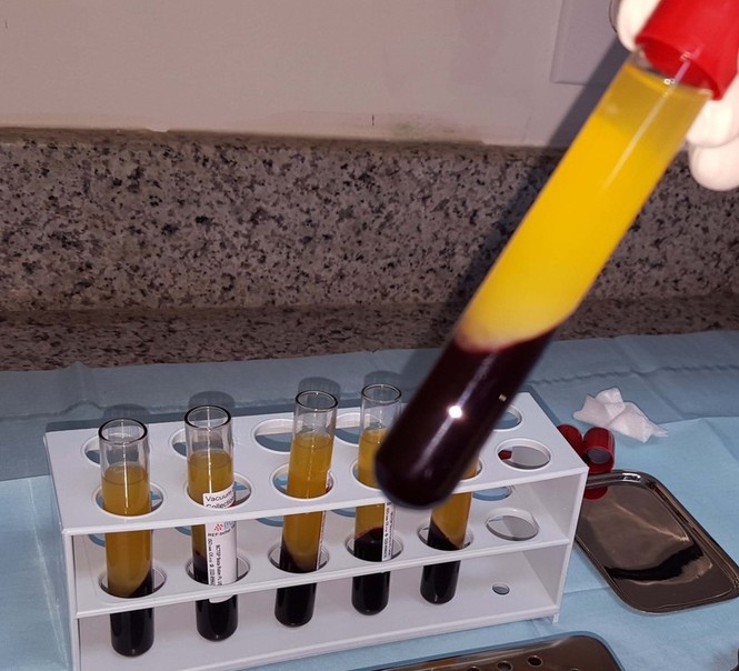
We need to return to the mechanisms of hemostasis. When tissue is injured, we obtain a blood clot and a fibrin clot. This fibrin clot releases growth factors from platelets and leukocytes. This promotes the formation of new blood vessels and stimulates cells, resulting in wound healing.
Platelets can secrete growth factors only after blood coagulation. This is why anticoagulants must be avoided in order to obtain a clot. This is the concept of PRF that I described in the early 2000s.
“PRF in Transplantology and Implantology,” Journal of Implantology, 2001.
It is possible to achieve physiological coagulation using biological products. Once centrifugation is complete, we obtain PRF, which consists of a blood clot and growth factors. Growth factors are proteins that stimulate cell differentiation, growth, and proliferation. They communicate with cells through specific receptors on the cell surface. Growth factors play a dominant role in wound healing. There are approximately 150 different growth factors:
- BMP — bone morphogenetic proteins
- FGF — fibroblast growth factor
- PDGF — platelet-derived growth factor
- IGF — insulin-like growth factor
- TGF — transforming growth factor
- VEGF — vascular endothelial growth factor, and so on.
Each of these factors has a specific function. For example, we want to understand why osteonecrosis of the jaw occurs when bisphosphonates are used. As we know, bisphosphonates alter bone metabolism. However, most importantly, their use critically reduces the secretion of keratinocyte growth factor (KGF). This factor is responsible for mucosal healing and keratinization.
This is why, following tooth extraction, we observe very slow healing and mucosal keratinization in these patients. Abnormal healing of the extraction site subsequently leads to bone infection and osteonecrosis.
So, which protocol should we use?
We know that PRP, PRGF, and PRF are available. If we take blood from the same donor into three tubes — PRP, PRGF, and PRF — each tube will contain the same number of platelets. The differences between these products result from the processing methods.
When preparing PRF, we do not use any processing additives. Why? Because we simply place the blood into a tube and allow it to coagulate physiologically. We do not need any additional agents. This makes the process more reproducible.
The type of coagulation affects the fibrin structure, and the fibrin structure determines its biological properties. If we need fibrin, it should be fibrin obtained through a physiological process. The healing outcome depends on the fibrin structure, fiber thickness, the number of networks formed, porosity, and permeability. This is why obtaining a natural, physiological fibrin clot is so important.
We can also compare these products based on their leukocyte content:
- PRP: 0–50%
- PRGF: 0%
- PRF: 50%
However, the most important property of platelet concentrates is how they release growth factors and how long this release continues.
- PRP — immediate release
- PRGF — unknown
- PRF — very slow release of growth factors, approximately 7 days or even longer.
Why is this property so important?
Fibrin acts as a carrier for these growth factors. Fibrin in PRF releases them slowly and continuously.
Research shows us how these factors can be effective. We need stable, continuous, and slow growth factor release to achieve good vascularization. We can compare this concept with biological processes.
For example, if we need to treat a very severe infection, what would we do? We would administer antibiotics through a slow intravenous infusion. The effectiveness of slow intravenous antibiotic infusion depends on maintaining a constant concentration.
Or consider a simple example: drip irrigation. Its principle is to supply small amounts of water continuously. This is why drip irrigation is considered one of the most effective methods. Drop by drop — that is the essence of irrigation.
Let us return to the PRF concept. As we know, blood collection must be performed quickly. If we do it slowly, drop by drop, the blood will begin to coagulate before we can separate the fibrin from the red blood cell mass.
After blood collection, we must immediately begin centrifugation, because we must remember that we are working without anticoagulants. If blood collection is performed slowly, we will obtain a poor-quality clot.
The only reason for failure is neglecting the timing. We must ensure that the tubes remain properly balanced in the centrifuge throughout the entire process. Balance is a very important factor.
Finally, we obtain natural PRF without contamination by red blood cells. PRF is located in the middle of the tube, between the platelet-poor plasma and the red blood cell mass. We can take this fibrin clot, place it on gauze, and use a special box to slowly and continuously compress it into a membrane.
This membrane can be used in a wide variety of clinical situations. If you need to divide it into small pieces, you can do so, unlike with an intact clot. You can also mix it with biomaterials or even suture it.
The procedure is very simple. We remove PRF from the tube, cut off the red blood cell mass, and place the clot into the box. Then we take the tray and apply pressure to obtain a membrane that is easy to use. The membrane will always have a certain thickness.
This is the result after a couple of minutes. The membrane is then very easy to manipulate. We can fold it in half and place it beneath the Schneiderian membrane during sinus augmentation. We can use a spatula to handle the membrane.
Alternatively, we can use the membrane to cover the biomaterial. A small amount of fluid remains at the bottom of the box. This fluid comes from the clot. Its main property is that it contains plasma and adhesive proteins, including fibronectin and vitronectin.
We can also create a perforated membrane by placing it on gauze and making incisions, as shown in the video. We make incisions in the gauze using a scalpel. We create as many perforations as necessary. We place the membrane on the gauze and make the holes in this way.
Then we return everything to the box. The membrane can be stored in the box for several hours, so it is important to return it to the box after handling.
We can then place it over the wound surface, for example, during immediate implant placement, when we want to cover the entire area with fibrin. In this case, the perforated membrane helps secure it around healing abutments or pins.
We know that flap elevation leads to bone resorption. To prevent this resorption, we use a PRF membrane.
As a result, we achieve increased alkaline phosphatase production, which reduces and prevents bone resorption. We can also protect the implant-abutment interface, the healing abutment, or the cover screw.
We also use a spatula to make membrane handling easier. You can see how easy it is to work with a PRF membrane.
We can also create a filler for socket preservation by mixing bone material with plasma under pressure. We place the clots into these cylinders, these tubes, and insert a special press. We must not apply excessive pressure, but at the same time, we must not apply too little compression.
This video was provided by our friend Dr. Polacci.
We place PRF into the white cylinders and then compress them. We stop at the level of the white cylinder. The press levels must be set correctly.
The first clot is placed easily. Subsequently, we obtain plugs that can be used to close extraction sockets. The more plugs we place into the socket, the better.
To compress these fibrin plugs, we must use gauze rather than a metal instrument. Each time, we gently compress the material using gauze.
Then we place guiding sutures to prevent the clots from falling out.

How did we begin?
We started in 2000 in my clinic. We treated an elderly woman with a severe infection of her leg. She was experiencing terrible pain.
I started using PRF not to reduce pain, but to stimulate new vascularization in the affected area. This is what we achieved after 10 days. We observed numerous blood vessels on the surface. And when we obtain blood vessels, we obtain healing. This is the essence of the concept: promoting vascular growth.
The next case involved complete amputation of the toes due to diabetes. After surgery, the patient developed an infection, and we treated it using PRF. The sole objective of this treatment was vascularization. After one week, we achieved healing. Here you can see the results at 7 days and 30 days.
Today, there is an enormous number of publications on PRF. We have sufficient evidence supporting its use not only in dentistry but also in orthopedics and aesthetic medicine.
However, if you ask me whether platelet concentrates always work, I will answer: no! Because in medicine, even if you have excellent materials, incorrect methods and protocols will lead to failure. Every protocol has its own clinical indications.
For example, here you can see PRF obtained using the correct technique. If we neglect the protocol during blood collection or centrifugation, we will obtain a poor-quality clot. Therefore, specific protocols must always be followed.
We must also use different protocols for different clinical situations. For example, PRF should always be placed beneath the Schneiderian membrane so that angiogenesis occurs from the mucosal side, because the Schneiderian membrane is a mucosal membrane. We must always follow the protocol.
For example, here the PRF membranes are folded in half. This gives us four layers. If we place two clots, each folded in half, we obtain four layers beneath the Schneiderian membrane.
In clinical practice, we know that flap elevation leads to bone resorption. When we raise a flap, we need to protect the surgical area to prevent bone resorption. The concept of PRF is that it acts as a tissue matrix and as a carrier for growth factors.
When we have a tissue deficiency, we can add a fibrin clot to the area. This fibrin will serve as an excellent matrix for the body to generate new tissue. This tissue can remain stable even after two years.
The concept of PRF is to deliver the fibrin matrix to the intended site. When we extract a tooth, we can fill the socket with biomaterial and cover it with fibrin. As we know, primary closure is not necessarily required.
Why? Because this position of the mucosa is physiological. If we respect biology, we should leave the mucosa in its physiological position. New mucosa will form in place of the fibrin matrix.
In this case, several teeth were extracted, as well as an implant. The dentist decided to use guided tissue regeneration with a Gore-Tex membrane.
The problem is that necrotic areas sometimes develop. In this case, primary closure will definitely not be possible, and the membrane should be removed. However, we know that fibrin can help us here. We cover the area with several layers of fibrin and simply leave it to heal.
Why is fibrin better in this situation? Because it is of natural origin. Unlike a collagen membrane or biomaterial, fibrin can be left exposed.
After 10 days, we can see blood vessels growing into the fibrin and new mucosa forming. After 8 weeks, new mucosa has formed in place of the fibrin clot.
If you want to assess the quality of the tissue, we can remove a small tissue sample for histological examination. However, we must first ensure that the graft is safe and that everything is functioning properly.
Here, the implantologist placed four implants. If we compare the mucosa on the right, which originated from fibrin, with the mucosa on the left, we see that it has a normal structure but is still immature and young.

Can we use PRF for root coverage as a connective tissue graft?
When we encounter gingival recession, we can use PRF as a matrix. However, the fibrin preparation protocol must be followed, and the fibrin must remain exposed externally.
Why? Because we want to achieve tissue gain. This is the essence of the matrix concept. Look at how the flap migrates upward into the area previously occupied by fibrin. As a result, we obtain well-keratinized tissue.
When we have an unfavorable gingival phenotype, particularly a thin phenotype, we can use PRF as a connective tissue graft. After flap elevation and tetracycline application, we place several layers of fibrin.
In this protocol, PRF also remains exposed and is positioned in front of the teeth. After one day, we can observe the formation of new blood vessels in the fibrin. If we achieve very rapid vascularization, we obtain tissue of excellent quality.
Here you can see keratinized tissue several months later. This is the concept of the fibrin tissue matrix. If we want to leave this matrix exposed, there is no problem, because the fibrin clot is of natural origin.
Several studies have compared PRF with connective tissue grafts. They obtained the same results as we did in our clinic. We can also use fibrin when harvesting a flap from the palate.
We know that this procedure is painful and healing is not always rapid. We suture fibrin into place and achieve faster healing in this area.
How should PRF be used for socket preservation after tooth extraction?
As I mentioned earlier, we should place as much PRF as possible into the socket while keeping the gingiva in its physiological position and avoiding tight closure of the socket edges.
We place sutures only to retain the fibrin in the socket. Then we obtain excellent healing. Guiding sutures alone are sufficient.
Whenever we perform an extraction, we usually try to use bone graft materials and collagen membranes. However, in my opinion, the best approach is to use only a fibrin clot.
We place a fibrin clot wherever extraction has been performed and then cover it with the flap. Look at how much bone has formed after three months.
But what matters most is not how much bone we have, but its quality. The quality of this bone is comparable to natural bone. It is completely natural bone.
We can then place implants into this viable bone. Of course, the implants will integrate well into such bone.
PRF in sinus augmentation
We can also use PRF in sinus augmentation. As I mentioned earlier, we fold the PRF membrane in two layers and place it beneath the Schneiderian membrane.
We can also use PRF to cover the created window, the defect remaining in the lateral wall. Today, we know that PRF can be used instead of a collagen membrane. There are publications supporting this technique.
We found no difference between using PRF and a collagen membrane to close the lateral window during an open sinus lift.
We can also use PRF to close perforations of the Schneiderian membrane. Fibrin adheres immediately to the mucosa and has excellent adhesive properties.
When perforations occur, we cover them with fibrin, perform the Valsalva maneuver, and continue the sinus augmentation procedure by filling the sinus with bone graft material.
Here you can see how the membrane heals, even though the test remains positive. We can also use fibrin as a biomaterial.

Today, several articles have been published on the use of PRF in lateral and crestal sinus augmentation techniques.
In the lateral sinus lift technique, implants are placed first, and then the sinus is filled with fibrin. After six months, complete regeneration of the sinus bone is observed. Therefore, fibrin is one of the best materials we can use for sinus augmentation.
Today, there are many publications on the use of PRF as the sole material for sinus filling.
I used this technique several years ago and followed this clinical case for nine years. The bone is very stable because it is entirely of natural origin.
When a perforation occurs, we close it with PRF, place the implants, and fill the sinus with a PRF clot. This is living bone, and it remains stable after six years.
Today, we also have several publications on this subject, including a recent one in the Journal of Craniofacial Surgery, describing the use of PRF as the sole material for sinus filling.
However, we practice medicine and surgery, and the most important thing is not the material. I can tell you that even if you use the best materials, you will not achieve good results if your protocols are incorrect.
It is extremely important to establish the correct indications. The indications depend on the palatal-to-buccal distance and the type of sinus.
When this space is narrow, I can guarantee you a 100% result. When the sinus is of medium width, we must carefully assess the situation. In wide sinuses, we should avoid using the PRF protocol.
When the space is narrow, we can fill it with fibrin, and the results will be fantastic. In narrow sinuses, we obtain bone because bone formation is easier in a confined space.
In wide sinuses, I ask you to forget this technique because you will not achieve the desired results. In such cases, only bone graft material should be used for augmentation.
PRF in immediate implant loading
We can also use PRF during immediate implant loading. When you perform extraction and implant placement and use bone graft material, the question always arises: how should we close the wound?
In this case, you can use fibrin because it is an excellent tissue matrix. Temporary crowns are then placed.
After one week, we obtain very good healing. If we remove the temporary crowns, we can see the newly formed mucosa. This is the tissue matrix concept.
After one month, we obtain an excellent aesthetic result. It is a very simple protocol.
Scientific evidence
Today, we can discuss the scientific evidence.
We need blood vessels because bone formation depends on the number of blood vessels. If we summarize our knowledge, we arrive at the following conclusion.
When we introduce bone graft material, we need cells because they produce the extracellular matrix. This extracellular matrix is necessary for blood vessel growth. And if we have blood vessels, we will have bone. Bone formation directly depends on vascular formation.
What is the essence of the PRF concept in bone formation?
PRF is active in bone formation because plasma proteins, such as fibrin and thrombospondin, act as precursors of the extracellular matrix.
First, we create the extracellular matrix. Then we achieve rapid vascularization and accelerated bone formation.
This is why I recommend using PRF mixed with biomaterial. First, cut PRF into small pieces using scissors. Then add the bone graft granules. We mix the granules with fibrin.
The mixture may be slightly dry, but this is not a problem because we can add the cellular fluid from the box. As a last resort, saline can be used if serum is unavailable.
Ultimately, we obtain an adhesive mixture of granules, fibrin, and serum. Using this protocol, the mixture becomes a sticky, plasticine-like mass.
PRF is active in hard tissues because it forms a highly organized fibrin matrix network deposited on extracellular matrix proteins such as type IV collagen or elastin.
PRF is capable of inducing vascularization in vivo. To moisten the biomaterial granules, we need extracellular matrix fluid. As we know, plasma proteins are very important for stimulating collagen gene expression.
There is a very interesting article comparing PRF clots and fibrin membranes. Should we use a fibrin clot? Never. We should use a membrane because it provides better vascularization — up to 100% more.
Advanced PRF — A-PRF
Now I can move on to the advanced PRF technique, A-PRF.
Why can we benefit from this development? Because scientists are obtaining new information about white blood cells. When we increase the number of white blood cells, we improve the results.
Monocytes are supercells in bone regeneration. Monocytes are very important as pre-osteogenic signals for progenitor cells. They induce the differentiation of osteoblasts from mesenchymal stem cells.
They also contain BMP-2 receptors, which means that we may be able to stimulate BMP production by these cells.
However, to obtain active monocytes, we must have active granulocytes because they are responsible for converting monocytes into macrophages. Therefore, monocytes and granulocytes must be incorporated into PRF without delay.
We developed new tubes. We modified the protocol and obtained 50% more VEGF. Because we use more white blood cells, we also obtain 20% more thrombospondin.
We obtain BMP-2 from the clot free of charge and for a prolonged period. We do not need to purchase it. We obtain a sufficient amount of PRF using advanced PRF and 50% more BMP-2-containing serum, which we can mix with the granules.
We can also coat the implant surface when necessary.
Scientific studies provide data on monocytes and macrophages. They undoubtedly produce BMP-2. We also observe a strong synergistic effect between monocytes and fibrin.
Fibrinogen enhances bone synthesis and the production of vascular-related factors by monocytes and macrophages.
If we compare PRF and A-PRF in vivo, A-PRF produces 2.5 times more blood vessels. This is achieved by increasing the number of white blood cells.
The concept of simple PRF is outdated today. It is not logical to exclude white blood cells. We need to induce inflammation, and inflammation will stimulate the formation of new blood vessels.

Clinical applications of A-PRF
Let us return to the clinical application of A-PRF.
We performed an extraction and left the extraction site without complete sutured closure. Why? Because we follow the biological concept, and this type of healing is physiological.
We should not attempt to achieve healing by primary intention. You can see the bone level and bone quality. We achieved complete ridge reconstruction.
When we perform this technique using A-PRF, we observe mucosal migration. There is no need to achieve primary closure.
Whenever we use this technique, we obtain natural wound closure within one week. According to this protocol, there is no need to use bone graft material because, after three months, bone will surround the implant without any bone graft material.
We can also use this concept when the buccal plate is missing. After filling the extraction site with A-PRF, we leave the wound to heal naturally. We can see that the patient’s own bone has formed after just three months.
When the buccal bone is absent, A-PRF is excellent for maintaining bone levels without biomaterials. By respecting biology, we leave the area open and achieve good results.
A-PRF also provides excellent results when used as a connective tissue graft, and these results may be even better than those achieved with conventional connective tissue graft techniques.
Alexander Alyam from Los Angeles performed excellent work using this protocol. Look at how we increased the amount of keratinized attached gingiva using PRF alone.
The technique is the same. We do not need connective tissue grafts or collagen membranes. We use only A-PRF.
Here you can see the healing process and the final result. Using PRF, we can obtain a good volume of keratinized tissue easily, quickly, and without pain.
Alexander Alyam further developed this concept to treat recession across an entire dental arch using only A-PRF — the tunnel technique.
In this case, eight membranes were used, all of them A-PRF membranes. This is the final result. We do not need to use any other products.

What are the future prospects for PRF?
Different techniques provide different degrees of PRF enrichment. The question is whether we need only platelets or something more.
We must look at blood itself. Platelets contain growth factors. We also need white blood cells. The presence of leukocytes is essential because they organize vascularization and osteogenesis.
Blood contains plasma, leukocytes, and platelets. We need blood containing white blood cells.
For this reason, we developed I-PRF. The “I” stands for injectable. This concept involves no additives and no anticoagulants. PRF is obtained automatically and is undoubtedly less expensive than PRP.
We need white blood cells to achieve vascularization. If we compare I-PRF with PRP and PRGF/AFG, we focus on the enormous number of white blood cells.
We obtain 20 times more than with PRP and more than 100 times more than with PRGF/AFG. We need monocytes, macrophages, and lymphocytes.
Look here: we have the same number of platelets. The I-PRF concept completely changes our understanding of PRF as a blood concentrate and specifically as a platelet concentrate.
This is what I-PRF looks like compared with PRP. We do not need a large volume of fluid. We need fluid containing a huge number of white blood cells because they are the cells responsible for promoting angiogenesis.
What do we want to achieve with I-PRF?
Sometimes we need to transform the gingival phenotype. If we inject I-PRF into tissue with a thin phenotype, we can change the characteristics of the tissue.
You can see how the number of blood vessels increased after two days and how vascularization improved. This case will be presented in Paris next month at a scientific symposium. This is injectable PRF.
We can also use I-PRF as a stabilizing biomaterial. Its stability is not the same as that of a bone block, but we can add I-PRF to bone graft material.
Look at what we obtain. We get a steak-like piece of biomaterial, which can subsequently be used as a bone graft. It provides the necessary stability to the bone granules.
We also obtain good protection for the granules because they are embedded in the fibrin clot. It is very easy to use and manipulate.
It takes only a couple of minutes to obtain this adhesive matrix, which can surround the bone graft. Naturally, we cover it with A-PRF and then perform mattress sutures.
Here is a case involving the mandible. We know that the mucosa in the mandible is highly mobile. In this situation, we can use a graft that is immobile and stable.
Creating this framework takes only 1–2 minutes. Then we cover everything with A-PRF membranes as usual and suture the area.
For information on how to prepare PRF clots correctly, read here.
Thank you for your attention. I hope this lecture has been useful. You can find more details on the website.
Video version of the PRF webinar: http://cross-dental.com/posts/1668894
If you are grateful for the information you have received, you may transfer any amount to the following bank details:
Ukraine: UA299358710000067321000114698 (FOP Verveiko V.I.)
Other countries: 4149500019820029 (Vitalii Verveiko)
PayPal: frankdent255@gmail.com


